| Concept Neuro & Spine Care hospital 24 Hour Emergency Facility Available |

Dr. Parimal Tripathi
 |
Dr. Ketan Patel M.S., M.Ch (Neurosurgery) |
Within the brain there are several ventricles, or cavities, that are filled with a clear liquid
called cerebrospinal fluid. The cerebrospinal fluid, which also surrounds the brain and
spinal cord, helps support and cushion the brain. Tumors in the ventricles are known as
intraventricular tumors, and they may arise from a variety of cells in the region. The tumors may
be astrocytomas, which arise from supporting cells in the brain; meningiomas, tumors of the
protective covering of the brain; ependymomas, which arise from the linings of the ventricles
themselves; colloid cysts and craniopharyngiomas, which arise from developmental cells; or
other brain tumors. As a whole, intraventricular tumors make up 10 percent of tumors in the
central nervous system.
Symptoms
When the flow of cerebrospinal fluid is blocked, it is a condition known as obstructive hydrocephalus. In people with hydrocephalus, the volume of fluid in the ventricle increases, placing pressure on surrounding brain tissue and leading to headache, nausea, mental status deterioration, visual disturbances, permanent neurological deficits, and death. Intraventricular tumors can cause other symptoms depending on location, including seizures, weakness or numbness in the limbs, impairments in language function, gradual changes in mood or personality, and memory loss. Imaging studies are the key component in the diagnosis of intraventricular tumors. Currently, magnetic resonance imaging (MRI) is the best available imaging modality. Computed tomography (CT) scans also are used, especially to assess hydrocephalus.
Treatment
Traditional treatment for intraventricular tumors often begins with the alleviation of the pressure caused by the obstruction in cerebrospinal fluid flow. Surgical tumor resection, the main treatment for brain tumors, can relieve the pressure as well as remove the obstruction. In some cases, the excess fluid must be drained off before surgery. This can be done with a shunt, an implantable tube that allows the excess fluid to drain to other parts of the brain or elsewhere in the body. Endoscopic surgery, in which instruments and cameras are manipulated through tubes inserted in a small incision, is useful for intraventricular tumors for several reasons. Cerebrospinal fluid is clear, making it easy for surgeons to visualize the tumor through an inserted camera. Also, because these tumors may arise from a wide variety of cells, a tissue biopsy often is necessary; with endoscopic surgery this can be done without open surgery. Finally, intraventricular tumors may be hard to reach with conventional surgical techniques, a restriction that does not apply to endoscopic surgery. A biopsy taken before or after surgery will be examined under a microscope to determine the tumor type and malignancy, and dictate follow-up treatment. Following surgery, patients may be treated with radiation therapy, chemotherapy, or both. In addition, some smaller tumors may be treated effectively with stereotactic radiosurgery, which involves the use of a highly focused beam of radiation to target the cancer cells specifically and leave the surrounding brain unaffected.
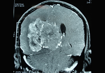
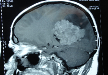
| Pre-operative | ||||
|---|---|---|---|---|
 |
 |
|||
 |
||||